


")


")


: Nominated for Drama Desk Award!")


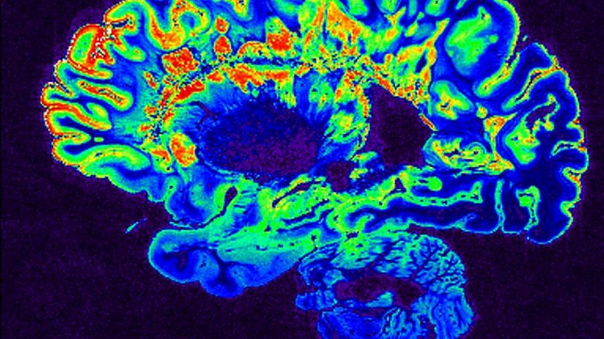
Researchers at the Hebrew University of Jerusalem discovered a way to transform a magnetic resonance imaging (MRI) machine into a device that can record changes in the biological makeup of brain tissue and help doctors determine earlier onset of diseases such as Alzheimer’s in order to begin treatment.
The findings were published on Tuesday in Nature Communications by Dr. Aviv Mezer and his team at HUJI’s Edmond and Lily Safra Center for Brain Sciences.
An aging brain or one that is affected by a developing neurodegenerative disease, such as Alzheimer’s or Parkinson’s, is marked by changes in the lipid and protein content of the brain tissue. Typical MRI scans do not detect these changes.
“MRI scans are already sensitive to these molecular changes, but until now, they were ignored, as all of the information comes in one block,” Mezer told The Times of Israel. “This [new tool] would provide physicians with a fuller picture of what is going on in the brain.”
Mezer added that the new model could pave the way for “doctors to compare brain scans taken over time from the same patient, and to differentiate between healthy and diseased brain tissue without resorting to invasive or dangerous procedures, such as brain-tissue biopsies that are currently done in cases of brain tumor.”
The new MRI technique will also provide more knowledge into how the brain ages, and the researchers believe that it can be also be used in the MRI of other body organs.
The model can already be applied to all MRI scanners.